Olympus BioScapes Competition
 Olympus Press Release
CENTER VALLEY, Pa., Dec. 11 /PRNewswire/—For most of us, red eye in our photos is nothing to be proud of. But for scientists around the globe, a brilliant crimson image of the inside of a mouse’s eye has set a new standard as one of the most beautiful and striking photos in the world. The image, captured by San Diego researcher Thomas Deerinck, triumphed over more than 1200 other entries to become the top prizewinner in the international 2006 Olympus BioScapes Digital Imaging Competition—the world’s foremost forum for showcasing microscope photos and videos of life science subjects.
Deerinck’s image was captured as part of research into neurofibromatosis, a usually fatal childhood illness that can cause tumors to form on the optic nerve and other locations in the nervous system. The image shows the optic fiber layer responsible for conducting information from the retina to the brain. Deerinck, who takes home Olympus products valued at $5000 as his prize, works at the National Center for Microscopy and Imaging Research at the University of California, San Diego.
Another key 2006 BioScapes Competition winner is a striking video depicting what has been described as the first-ever visualization of cytotoxic T-Cells zeroing in on target cells and killing them. The video was captured by Dr. Thorsten Mempel of Harvard University (Fifth Prize). Still other fascinating and beautiful images depict the muscles that make the singing toadfish “sing,” cervical cancer cells, a new insect species, a human fingertip, a variety of flowers and plants, and a close-up showing how a wound in living tissue begins to heal itself.
The Olympus BioScapes competition is the world’s premier platform for honoring images and videos of plant, animal and human subjects as captured through light microscopes. Any life science subject is eligible, and entries are judged based on the science they depict, their aesthetics (beauty and impact of the image), and their technical merit. This year, in addition to Prizes 1-10, 54 other images and movies were recognized with honorable mentions. All images and the names of all honorees may be viewed online at www.olympusbioscapes.com.
Winners of this year’s competition were recognized last night at San Diego’s elegant Horton Grand Hotel; 21 of the images will be on display from December 12, 2006 to March 5, 2007 at the San Diego Natural History Museum. After leaving San Diego, the photographs will tour museums throughout the U.S. as part of a program developed in tandem with Natural History Magazine. Tour stops include Los Angeles, Woods Hole, Mass., Ann Arbor, Mich., New York City and Philadelphia. Additional BioScapes exhibits will tour St. Louis, Allentown, Penn., Dallas and suburban Washington, D.C.
“These images and videos document scientific events that give us insight into the most important aspects of nature,” said Stephen Tang, Ph.D., Group Vice President and General Manager, Life Science, for Olympus America. “But they also tell stories of great meaning and emotion. It is one thing to hear about a disease; it is entirely another to see an extraordinary photo of great beauty and power, and then realize with a shock that you are looking at cancer cells. Similarly, seeing this year’s movie of T-cells targeting cells and killing them is like watching a thrilling adventure film.”
Every year, Olympus selects four outstanding authorities in microscope imaging as its panel of judges for the noncommercial competition, which is open to users of any brand of light microscope and camera equipment. This year’s BioScapes judges included Dr. Douglas Murphy, Director of the Light Microscopy Facility at the Howard Hughes Medical Institute’s Janelia Farms Research Center in Virginia; Dr. Simon Watkins, Professor of Cell Biology, Physiology and Immunology, and Director of the Center for Biologic Imaging, at the University of Pittsburgh; Dr. Sue Aicher, Associate Professor at the Neurological Sciences Institute of the Oregon Health & Science University; and Dr. Kenneth N. Fish, Assistant Professor in the Department of Psychiatry, University of Pittsburgh.
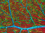
In addition to Thomas Deerinck and Thorsten Mempel, other Top 10 winners included: Hermann Aberle of MPI for Developmental Biology, Tuebingen, Germany, for his image of fruit fly muscles and axons; M. R. Dadpour of the University of Tabriz, Iran, for his photo of a zinnia flower; Robert Markus of the Hungarian Academy of Sciences, Csongrad, Hungary, for his Mirabilis jalapa stamen; Ron Oldfield of Macquarie University, Sydney, Australia, for his photo of Dictyostelium sp. (slime mold); Wim van Egmond of Rotterdam, The Netherlands, for his Dinobryon (golden algae) image; Ralph Grimm of Jimboomba, Australia, who captured the proboscis of a common housefly; of Charles University, Prague, Czech Republic, for his Cynoglossum officinale image; and Michael Janes of Invitrogen Corp., Eugene, Oregon, for his photo of bovine pulmonary artery cells.
To view all the winning images and see a complete list of the winners and honorable mentions, visit www.olympusbioscapes.com . For free access to the images, media members and other noncommercial users may contact [email protected].
About Olympus
Olympus is a precision technology leader, creating innovative opto-digital solutions in healthcare, life science and consumer electronics products. Olympus works collaboratively with its customers and its parent company, Tokyo-based Olympus Corporation, to leverage R&D investment in precision technology and manufacturing processes across diverse business lines. For more information, visit www.olympusamerica.com. For information on the Olympus BioScapes Digital Imaging Competition, visit www.olympusbioscapes.com.